
























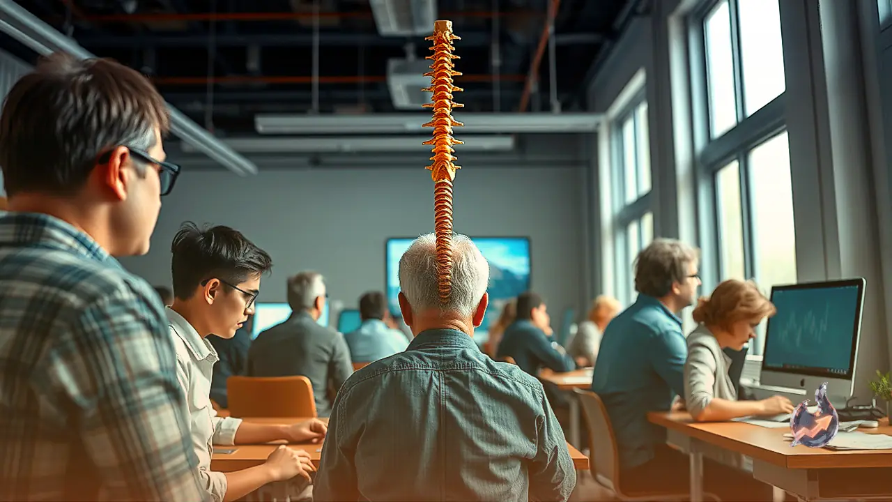

Serving as the structural foundation for human posture and bipedal mobility, the spinal column extends from the base of the skull all the way to the pelvis through a sequence of interlocking vertebrae. This intricate biological pillar comprises three principal segments. At the apex lies the cervical spine, consisting of seven vertebrae that support cranial movement. Immediately below, extending along the ribcage, rests the thoracic region, which incorporates twelve vertebrae. The lower back is supported by five robust lumbar vertebrae, underneath which five fused sacral bones form a solid pelvic anchor, terminating in the coccyx or tailbone. Running through the center of these stacked skeletal rings is a hollow tubular canal that houses the spinal cord. From this central channel, a vast network of neural pathways branches outward to innervate the entire body, delivering sensory information to the brain and transmitting operational commands back to peripheral tissues.
The entire physical load of the torso relies upon this bony architecture, while the neural conduits within mediate voluntary and involuntary bodily functions. Under contemporary lifestyle demands, individuals aged twenty to thirty are displaying an alarming decline in spinal integrity. Young adults frequently exhibit structural deterioration and functional complaints once restricted solely to geriatric age groups. Two major lifestyle patterns drive this physiological crisis: mechanical stress from screen engagement, commonly termed tech neck, and prolonged physical immobility, characterized as a sitting epidemic. Examining the biomechanical degradation behind these factors reveals the extent of their systemic impact.
Biomechanical Load on the Inclined Neck and Structural Flattening
Under neutral alignment, where an individual looks straight ahead without vertical displacement, the head exerts a gravitational force of approximately 4.5 to 5.5 kilograms on the cervical joints. In this balanced zero-degree configuration, structural equilibrium is preserved. However, when an individual tilts their head downward to an angle of 60 degrees to browse a handheld display, the effective mechanical force borne by the cervical vertebrae escalates sharply, adding an extra 27 kilograms of continuous tension. This persistent mechanical overburden triggers repetitive strain throughout the dorsal region.
Enduring such disproportionate biomechanical resistance subjects surrounding muscular fibers, stabilizing ligaments, and connective tendons to continuous micro-trauma, precipitating chronic localized inflammation. Over time, the sustained anterior tension alters the natural structural curvature of the cervical spine, which normally features a gentle lordotic C-shaped arc. As the curve flattens into an unnatural vertical alignment, the condition known as cervical lordosis takes hold. This skeletal displacement disrupts cervical load distribution, destabilizes tendon anchors, compresses traversing nerve roots, and drastically accelerates premature intervertebral disc degeneration and bulging.
The Metabolic and Musculoskeletal Toll of Constant Desk Confinement
Sedentary confinement represents a pervasive behavioral challenge of modern work cultures, wherein working professionals and students routinely remain seated for eight to twelve hours each day. Daily routines involve stationary desk seating during working shifts, continued chair immobility during study intervals, and reclined screen engagement during leisure periods. Operating digital devices while lying down creates particularly chaotic strain angles across the upper spine, confounding natural ergonomics.
Maintaining an uninterrupted seated posture directs the cumulative force of gravity straight onto the lumbar spine and pelvic basin. Consequently, the stabilizing musculature of the lower back turns dormant, losing both tone and contractile readiness. This lack of dynamic movement decelerates systemic circulation, diminishes muscle elasticity, and places uneven, asymmetrical pressure upon the lower intervertebral cushions. As a result, the protective discs experience accelerated mechanical wear. Furthermore, the sharp downturn in metabolic rates contributes to secondary pathologies, including adiposity, systemic insulin resistance, and an early-onset epidemic of chronic vertebral degeneration.
From Disc Herniation to Neural Deficits and Loss of Motor Function
From an orthopedic perspective, habitual sitting and prolonged cervical flexion represent overlapping facets of ergonomic neglect. When persistent muscular stiffness and neck soreness are dismissed as inconsequential fatigue, mechanical wear shifts toward irreversible neurological pathology. Between adjacent vertebral segments lie fluid-filled cartilaginous pads known as intervertebral discs, which serve as shock absorbers and permit flexibility.
Sustained abnormal alignment and muscular spasms gradually dehydrate this inner gelatinous nucleus, stripping the disc of its shock-absorbing capacity. Eventually, the weakened outer ring tears or bulges outward from its designated boundary, culminating in disc herniation or a slipped disc. This structural rupture generates intense localized discomfort. Because vital neural roots originating in the cerebral center traverse through this cervical junction to reach the arms and legs, persistent focal compression impedes signal transmission, inducing sensory numbness across the extremities.
Motor Failure and Severe Loss of Autonomic Control
Should structural compression continue unabated, the deficit progresses from sensory paresthesia to profound motor deterioration. Patients discover that even with deliberate conscious effort, their muscular groups fail to generate sufficient mechanical torque. Manual dexterity degrades, grip strength diminishes, and everyday objects slip unintentionally from the fingers.
As lower spinal pathways become compromised, neuromuscular coordination in the lower limbs begins to falter, resulting in an unsteady, staggered gait. In advanced, critical stages where the spinal canal experiences extreme narrowing and central cord impingement, control over autonomic functions can disintegrate, leading to the loss of voluntary bowel and bladder continence. Such neurogenic sphincter disruption constitutes an acute orthopedic and neurological emergency demanding urgent medical intervention. Immediate posture correction and the mitigation of constant screen flexion remain indispensable to ward off permanent structural paralysis.