

















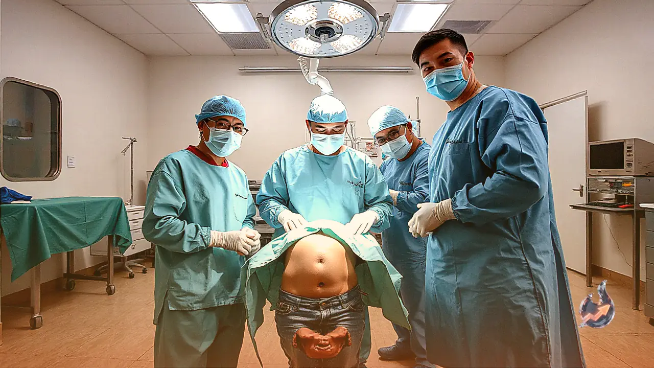

A surgical team at New Medical College Hospital in Kota, Rajasthan, has successfully performed a complex, high-risk surgical procedure, granting a teenage girl a fresh lease on life. The young patient had been enduring severe abdominal distress for an extended duration. Upon conducting advanced clinical evaluations, medical specialists discovered hundreds of varying-sized fluid-filled cysts distributed across her abdominal cavity. The surgical department mobilized immediately, performing an extensive operation to excise the lesions, which collectively weighed approximately 10 kilograms.
According to operating surgeons, the cysts numbered between 400 and 500 individual sacs. The massive parasitic growth occupied nearly her entire abdomen, asserting intense pressure against vital internal organs and triggering chronic, debilitating pain. During the surgical intervention, several cysts ruptured, discharging their fluid into the cavity. Surgeons carefully evacuated the leaked contents, thoroughly cleansed the region to prevent secondary complications, and completed the removal without harm to adjacent structures. Following the procedure, the girl remains under observation and is recovering steadily in stable condition.
Initial local clinic visits failed to provide relief
The patient had been experiencing recurrent abdominal pain over an extended timeframe. Her family initially sought medical consultation at a local peripheral health facility, where she received standard therapeutic courses. Despite undergoing medical care there, her condition showed zero improvement, and the severe symptoms persisted unabated. Recognizing the escalating severity of her discomfort, her relatives transferred her to New Medical College Hospital in Kota for tertiary evaluation.
She was admitted directly under Unit C of the hospital's Department of Surgery, where senior specialists initiated an intensive investigative protocol. Given her prolonged distress and visible abdominal swelling, clinicians immediately arranged ultrasonography and comprehensive CT scans. The diagnostic imaging revealed extensive collections of fluid-filled cystic structures dominating her abdominal cavity. Confronted with the massive burden and the clear hazard of organ compromise, the surgical team decided on prompt surgical intervention.
Abdominal cavity packed with hundreds of membrane-bound sacs
Upon entering the operative field, the surgical team encountered an extraordinary clustering of small and large cysts dispersed throughout the peritoneal cavity. Ranging across nearly the entire abdominal space, the 400 to 500 cysts were directly compressing surrounding organs, severely inhibiting normal physiological functions. The aggregate mass of the extracted fluid sacs stood at approximately 10 kilograms.
Surgeons observed that each cyst possessed a distinct, skin-like outer capsule encasing thick fluid within. The physical mass and high internal pressure caused several individual cysts to burst spontaneously during the intricate dissection, spilling their liquid contents into the abdominal space. The surgical team acted methodically to aspirate all released fluid, carefully separating the remaining intact cysts from delicate visceral tissue before extracting the entire cluster to avoid systemic toxicity.
Suspected parasitic infection linked to infected canine waste
Detailing the clinical etiology, Dr. RG Meena explained that such conditions typically stem from a parasitic worm that can enter the human gastrointestinal tract through inadvertent exposure to infected dog feces. Once established internally, the parasite multiplies and prompts the gradual formation of multiple fluid-filled cysts within visceral organs and surrounding cavities. Presentations of this magnitude, featuring hundreds of distinct cysts, are considered exceptionally rare in routine surgical practice.
To definitively ascertain the precise pathogen and cellular structure of the lesions, surgical staff preserved representative tissue samples and submitted the excised cysts to the Department of Histopathology for thorough microscopic analysis. While the clinical pattern strongly aligns with an advanced parasitic infestation, formal confirmation of the causative agent will depend on the final histopathological findings.
Operation led by Dr. RG Meena and surgical specialists
The intricate surgical procedure was spearheaded by Dr. RG Meena, Professor and Head of Unit C in the Department of Surgery, supported by a multidisciplinary team of medical professionals. Beyond the technical challenges of the excision, the medical unit maintained vigilant post-operative monitoring to stabilize the patient, track vital signs, and prevent post-surgical infections.
Hospital officials and the operating team consider the successful outcome a major institutional milestone. Clinicians noted that left unoperated, the expanding 10 kg mass would have inevitably caused critical organ failure or life-threatening systemic shock. With the girl now recovering well, physicians await the histopathology report to determine whether targeted anti-parasitic pharmaceutical therapy will be required during her post-operative care.