
























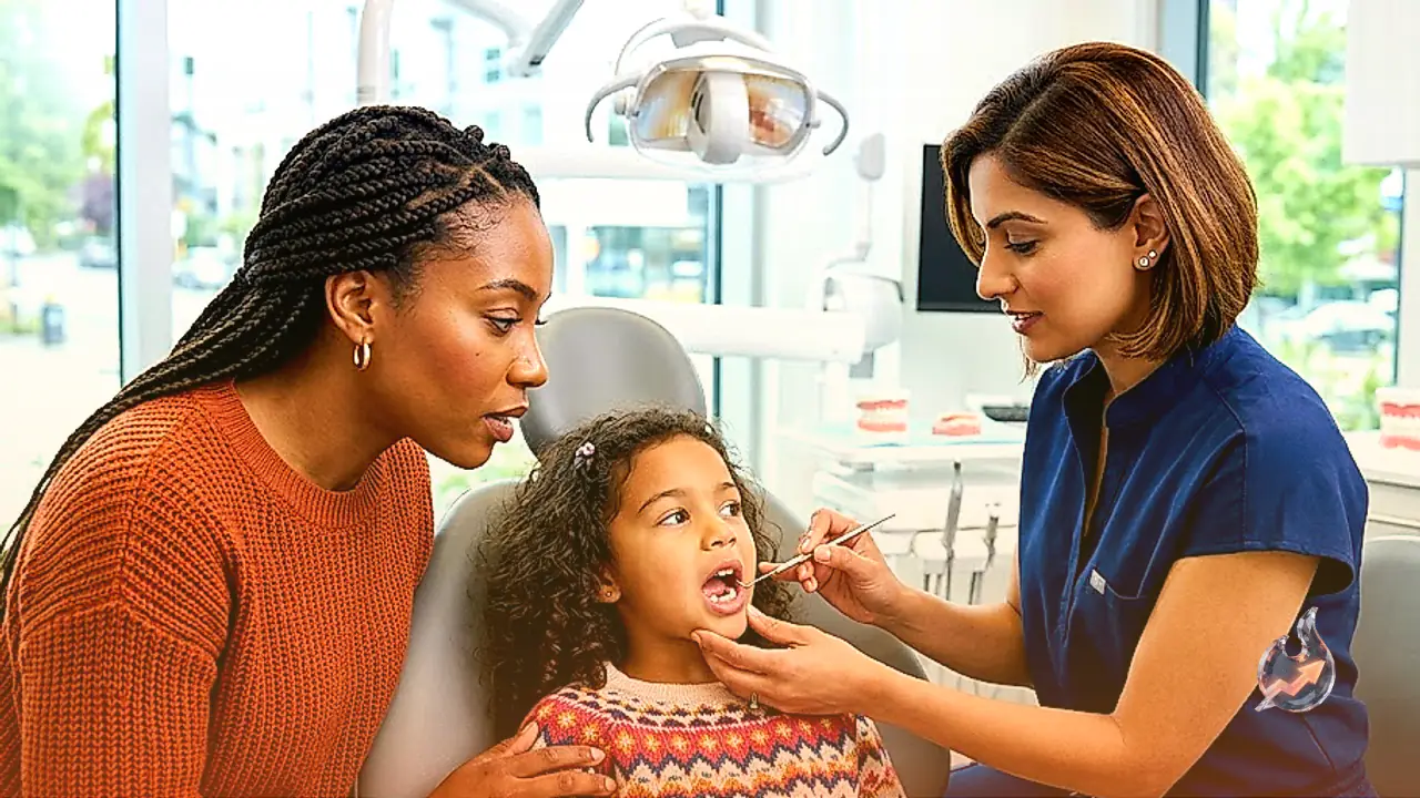

During a child's natural physical development, the shedding of primary teeth follows a predictable timeline where milk teeth gradually loosen and yield their positions to strong permanent teeth. Nevertheless, unexpected falls during play, sudden physical injuries, or aggressive cavity progression frequently cause primary teeth to detach far ahead of schedule. When an empty socket appears prematurely, parents routinely worry about the enduring consequences this gap might impose on the emergence of incoming adult dentition. A primary concern revolves around whether surrounding teeth will drift into the newly formed vacuum, ultimately compelling the growing child to endure lengthy orthodontic alignment therapies later in life. Pediatric dental specialist Dr. Megha Jain, from the dental department at Amrita Hospital in Faridabad, explains that primary teeth play an indispensable role in safeguarding oral architecture.
The function of primary dentition extends well beyond the basic mechanics of chewing meals and developing speech articulation. In clinical reality, these initial teeth serve as indispensable anatomical pathfinders for the adult teeth taking shape deep inside the gum tissue. When a milk tooth remains securely anchored until its natural biological shedding phase, it reserves the necessary horizontal space and creates a directional guide path for the adult tooth erupting underneath. If this biological spacer is knocked loose or extracted prematurely, the underlying permanent tooth loses its navigational marker, increasing the likelihood of an impeded or aberrant eruption path.
Severe Cavities and Infections Weaken Tooth Anchoring
Discerning whether a child has shed a milk tooth naturally according to age or lost it prematurely is nearly impossible through casual visual inspection at home. A precise diagnosis requires professional evaluation utilizing specialized pediatric examination tools. In numerous pediatric cases, bacterial decay advances unchecked, severely compromising the structural integrity of the enamel and dentin. When microbial invasion reaches the root canal system, purulent discharge and localized abscesses can develop around the surrounding gum tissue. Such aggressive infectious conditions undermine the periodontal ligament and bone support, causing the tooth to loosen and fall out long before its physiological lifespan concludes. Recognizing any persistent discoloration, swelling, or foul odor warrants an immediate clinical assessment.
Surrounding Teeth Migrate Toward the Empty Space
Human teeth do not exist in absolute rigid isolation; rather, they react dynamically to adjacent forces and chewing pressures within the dental arch. The moment a primary tooth exits prematurely and leaves a vacant socket behind, the equilibrium of the dental arch is disrupted, prompting nearby teeth to migrate toward the void. Posterior teeth naturally tilt and drift forward into the gap, while anterior teeth may shift backward into the opening. This progressive drift steadily narrows the corridor required by the underlying adult tooth. Consequently, when the permanent successor finally attempts to erupt, the restricted arch space frequently forces it into an irregular angle, causing crowding or malposition. In severe cases, the child may subsequently require comprehensive orthodontic interventions and braces to restore normal alignment.
Diagnostic Radiographs Govern Space Maintainer Usage
Not every pediatric patient who experiences early tooth loss automatically requires an artificial appliance known as a space maintainer. The clinical decision to insert this prosthetic spacer depends on a meticulous evaluation of multiple developmental parameters. Specialists carefully examine the precise chronological age of the child and identify the specific tooth type that has been lost prematurely. Concurrently, clinicians must determine the developmental stage and eruption readiness of the permanent tooth maturing beneath the gum line. Specialized dental radiographs are conducted to visualize the precise dimensional gap separating the vacant crest from the underlying adult tooth crown. Only after evaluating these imaging scans will dental specialists confirm whether an appliance is essential to preserve the arch perimeter.
Essential Safeguards Following Traumatic Oral Injuries
When physical impact or athletic accidents knock out or fracture a primary tooth, guardians should escort the child to a dental professional without delay. Caregivers must observe a strict medical precaution: never attempt to manually reinsert an avulsed milk tooth back into the bleeding socket at home, as forceful manipulation risks damaging the sensitive germ of the permanent tooth residing below. Instead, the dislodged tooth can be wrapped in a clean, sterile cloth and transported directly to the clinic for inspection. During emergency clinical evaluations, practitioners examine far more than the empty space, conducting thorough inspections of the gums, inner lips, neighboring dental units, and underlying alveolar bone for unseen fractures.
In the aftermath of oral trauma, young children commonly experience localized discomfort, tenderness, and irritation across the wounded gingival tissue. Caregivers should actively discourage the child from touching the vulnerable socket with their fingers, because introducing external pathogens dramatically heightens the risk of secondary infections. If intense pain, persistent swelling, or abnormal bleeding continues unabated, prompt re-evaluation by a specialist is vital. Timely clinical intervention remains the most reliable strategy for preserving dental arch integrity and safeguarding the pathway for upcoming adult teeth.