















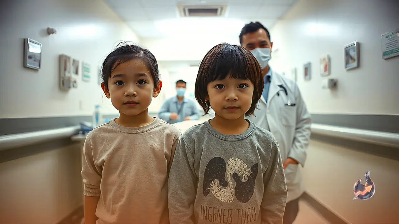

A rare and startling medical anomaly has come to light in the Pali district of Rajasthan, where medical specialists were astonished during a diagnostic workup of two siblings who arrived seeking treatment for persistent respiratory troubles. While human anatomy typically places the heart on the left side of the chest and the liver on the right side of the abdomen, detailed scans revealed that the internal vital organs of both children are positioned in complete reverse orientation, behaving like a mirror image of normal human anatomy.
Routine Examination Leads to Astonishing Diagnostic Findings
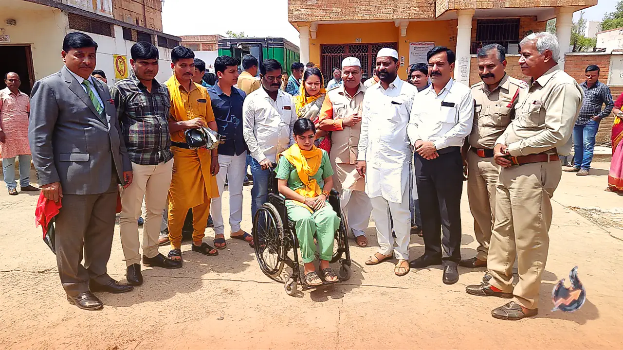
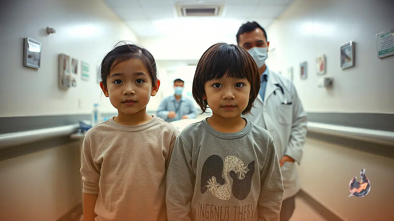
According to Dr. Laxman Soni, a respiratory medicine specialist at Bangur Hospital in Pali, family members brought a 13-year-old girl and her 10-year-old brother to the outpatient facility on October 25. The parents explained that both children had been struggling with unremitting cough, laboured breathing, and recurrent episodes of pneumonia since their early infancy. Noting the chronicity and severity of their chest symptoms, the medical team ordered a comprehensive battery of diagnostic evaluations.
The investigative protocol included high-resolution computed tomography scans, abdominal ultrasound sonography, echocardiography, and other supportive investigations. The clinical team was taken aback when reviewing the radiographic films, as the results conclusively demonstrated that both the boy and the girl had their hearts situated on the right side of their thoracic cavities, while their livers occupied the left side of their upper abdomen. Encountering such identical anatomical reversals simultaneously in two siblings from the same household is an extraordinary occurrence in clinical practice.
Understanding Situs Inversus and Kartagener Syndrome
In medical terminology, the complete anatomical reversal of internal thoracic and abdominal viscera is designated as situs inversus. This uncommon presentation is estimated to occur in roughly one out of every 50,000 live births, representing a fully congenital and genetically linked condition. The broader clinical spectrum is often classified under Primary Ciliary Dyskinesia (PCD) and Kartagener syndrome, wherein structural abnormalities affect the functioning of microscopic hair-like organelles known as cilia.
Under normal physiological conditions, these microscopic cilia lining the respiratory epithelium beat rhythmically to clear inhaled debris, environmental dust particles, and excess mucus out of the airways. In individuals affected by PCD or Kartagener syndrome, the cilia are either completely immobile or move dyskinetically, resulting in the chronic stagnation of respiratory secretions. Consequently, individuals living with this condition endure a persistent cough, sinus trouble, and frequent, severe lower respiratory tract infections throughout their lives.
Why Occurrence in Multiple Siblings Is Medically Significant
Medical experts emphasize that the simultaneous presence of congenital organ transposition and chronic respiratory symptoms in two biological siblings makes this observation remarkably noteworthy. Multiple forms of PCD are transmitted from parents to offspring through autosomal recessive inheritance pathways. Nevertheless, clinical specialists point out that an anatomical transposition alone does not constitute complete definitive confirmation of the primary molecular defect; comprehensive genetic testing is essential to confirm the precise mutational profile.
Physicians advise parents and guardians never to disregard persistent or recurring coughs in young children as mere seasonal colds. Seeking timely consultations with pulmonary specialists and obtaining appropriate radiologic evaluations can ensure an early and accurate diagnosis, thereby preventing long-term irreversible lung damage through structured medical management.